To the average cat owner, cardiomyopathy may sound like a scary mouthful of a word — but all it essentially means is disease of the heart muscle. And cardiomyopathy is not one single disorder, but instead a family of heart conditions that can be classified into three types: hypertrophic cardiomyopathy (HCM); dilated cardiomyopathy (DCM); and restrictive cardiomyopathy (RCM).

288
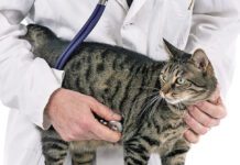
The causes, signs and treatments are different for each of the conditions, and rate of progression and prognosis also vary between individual cats and between breeds. “This is yet another important reason for the regular veterinary examination,” explains Marc S. Kraus, DVM, senior lecturer in the Section of Cardiology at Cornell University’s College of Veterinary Medicine. “The exam will enable the veterinarian to feel any pulses in the hind legs, listen for a murmur or an arrhythmia in the heart, and also listen to lung sounds. The veterinarian will be able to determine if an early form of cardiomyopathy is present — and this is when the condition is most manageable.”
While occasionally observed in kittens, cardiomyopathy is almost always an acquired condition and is by far the most common among all adult feline heart disorders — accounting for almost two-thirds of heart conditions diagnosed in cats. Cardiomyopathy is brought about by a structural abnormality in the muscle enclosing one or both ventricles, with the affected chamber taking on a thickened, dilated or scarred appearance. (The left ventricle is almost always affected; right-chamber involvement may also occur, but only rarely.)
This abnormality sets the organ’s blood-collecting and blood-pumping mechanics amiss, and this dysfunction can progress to congestive heart failure — and a resulting collection of fluid in or around the lungs — and then to respiratory distress. Other potential outcomes of cardiomyopathy include paralysis-causing blood clots, which usually arise from the left atrium and lodge in arteries somewhere in the body — usually those supplying the rear limbs. Arrhythmias, kidney disease, and sudden death can also occur.
Most feline cardiomyopathies are considered to be primary diseases, ie. those whose origins are either genetic or unknown. However, some are secondary diseases — those whose causes are specifically identifiable and include such conditions as anemia, hyperthyroidism and high blood pressure.
Hypertrophic Cardiomyopathy. HCM is the most common of the cardiomyopathy classes, and is also the most likely to be detected during the annual checkup of an outwardly healthy feline patient. General signs include poor appetite, weakness and sometimes vomiting, but it is also not unusual for the cat to seem quite normal at home until the heart goes into advanced failure.
This disorder results in the thickening of one or more parts of the heart muscle, leading to changes in the way the heart pumps blood. For whatever reason, certain breeds — Main Coon, American Shorthair, Persian and Ragdoll cats — have an increased risk of this condition. In the Main Coon, HCM is considered to be an inherited disorder, and it is strongly suspected to be inherited in the other breeds, as well. (See related sidebar on next page.)
The onset of the disease is typically during young adulthood, with males more frequently afflicted than females. However, cats can be very young (especially in Ragdolls) or well into adulthood (10 years old) when the condition is initially diagnosed. Murmurs or gallop rhythms may occur, or no clinical signs may be noted. Unfortunately, heart failure can occur quickly once the heart becomes affected, and fluid buildup in the chest cavity can make breathing very difficult. These cats may also develop blood clots in the arteries, especially in those that supply the hind limbs.
Dilated Cardiomyopathy. It was once a common condition, but modern dietary formulations with higher levels of taurine have led to a significant drop in disease rates. This condition leads to a thinning of the muscle wall of the heart, resulting in weak pumping and a reduced forward flow of blood from the heart. The diagnosis is usually made in slightly older cats that are showing signs of heart failure.
A slow heart rate, low blood pressure and low body temperature may be noted, and these cats may also form blood clots in the arteries. There may be gallop rhythms or murmurs noted by the vet. These days, DCM is seen mostly in cats fed dog food or homemade diets without appropriate taurine supplementation.
Restrictive Cardiomyopathy. In this disease, the heart becomes constricted in its pumping ability due to loss of elasticity, which makes the heart function in a stiff manner. Scar tissue or inflammation of the muscle may be responsible for these changes. Rhythm disturbances and murmurs may be detected, and blood clots may get lodged in the arteries. It most often affects geriatric cats.
Diagnosis and Treatment. This is usually accomplished with a chest radiograph (X-ray) by your cat’s veterinarian, and the heart will have an abnormal shape. Fluid may be detected in or around the lungs. However, if a large amount of fluid is present around the lungs, it may be necessary to remove the fluid first and take more radiographs to more accurately evaluate the heart.
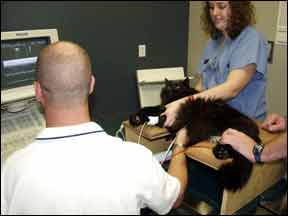
Ultimately, definitive diagnosis requires an echocardiogram (or sonogram), a non-invasive method of looking at the heart while it is pumping. Sound waves are used to make this dynamic study of the heart. Radiographs can tell us about the size and shape of the heart but nothing about heart function; however, ultrasound can provide us with this information. Ultrasound will also allow measurement of the heart muscle to determine if it is too thick (hypertrophic or restrictive CM) or too thin (dilated CM). Finally, an electrocardiogram (EKG) is useful to evaluate the rhythm of the heart.
Treatment will be based on the type of cardiomyopathy present, and tailored to the needs of the individual pet. Different drugs are used for the different forms. Supporting the strength of heart contractions, reducing fluid buildup, nursing and supportive care and dietary adjustments (such as a low sodium diet) are some of the common strategies a vet will attempt.
Therefore, if at all possible, tests necessary to define the specific forms are performed before treatment begins. Fortunately, most of these cats can be stabilized with the correct drug; however, continual medication may be necessary since the disease cannot be cured.
Determination of the level of thyroid hormone (T4) in the blood is also sometimes beneficial in evaluating cats with hypertrophic CM. This simple blood test can help identify an overactive thyroid gland as the underlying cause of the heart disease. This form of CM is potentially reversible if the cat receives appropriate and timely treatment for the thyroid disease.
Complications To Beware. Most of the cats with cardiomyopathy develop signs of heart failure as previously described; however, these cats are also prone to producing blood clots within their hearts. When these clots escape the heart, they travel through various arteries leading from the heart and eventually lodge in a narrow part of the artery. The most common site for clots to lodge is the point at which the aorta splits before going into the rear legs.
Therefore, these cats often become paralyzed very suddenly and are in significant pain. In many cases, it is paralysis and pain that first becomes noticeable and is the reason that medical treatment is sought. This may be mistaken for an uncomplicated lameness or even a broken leg. Within a few minutes to hours, there are no pulses in one or both rear legs, the legs are cold and the footpads appear blue due to the lack of oxygen.
“Sometimes the animal can’t walk in either their hind limbs or their front limbs, although a saddle thrombosis [weakness or total paralysis in the hind limbs] is most common,” explains Dr. Kraus, who is board-certified by the American College of Veterinary Internal Medicine (Cardiology). “A clot can go into the lungs, or the intestinal tract, which will create excruciating pain for the animal. The cat will display lethargy, not wanting to eat or walk. The most common sign is that they can’t move, or they will retreat under the bed and won’t come out.”
Treatment of the cat concentrates on drugs to relieve pain and to hasten the return of circulation to the legs. Since these cats also have severe heart disease, they make poor surgical candidates. Therefore, surgery to remove the clot is not advisable due to the high incidence of death during surgery. Medical therapies with “clot buster” drugs have been associated with a high degree of morbidity and mortality.
The prognosis for CM is variable depending on the form of the disease and the severity at the time of diagnosis. Many cats will live years if properly medicated. If congestive heart failure is already present when cardiomyopathy is detected, the survival rate declines with an average of three to 12 months.
If the disease is detected in its asymptomatic state however, your cat may live a long life with close monitoring of the condition by your veterinarian or a veterinary cardiologist. The exception is when CM is caused by hyperthyroidism. If hyperthyroidism is successfully treated, the heart function will generally return to normal and the cat will no longer require treatment.
“It’s important for the pet owner not to panic,” stresses Dr. Kraus. “Many cats with cardiomyopathy are not being dealt a death sentence. If they’re not in heart failure, the animal can live happily for many years before developing full-blown cardiomyopathy. It all depends on where the disease process is when it’s diagnosed.”


