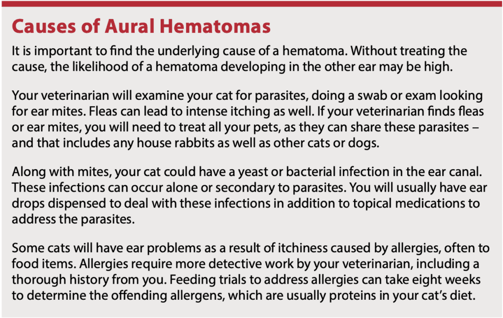
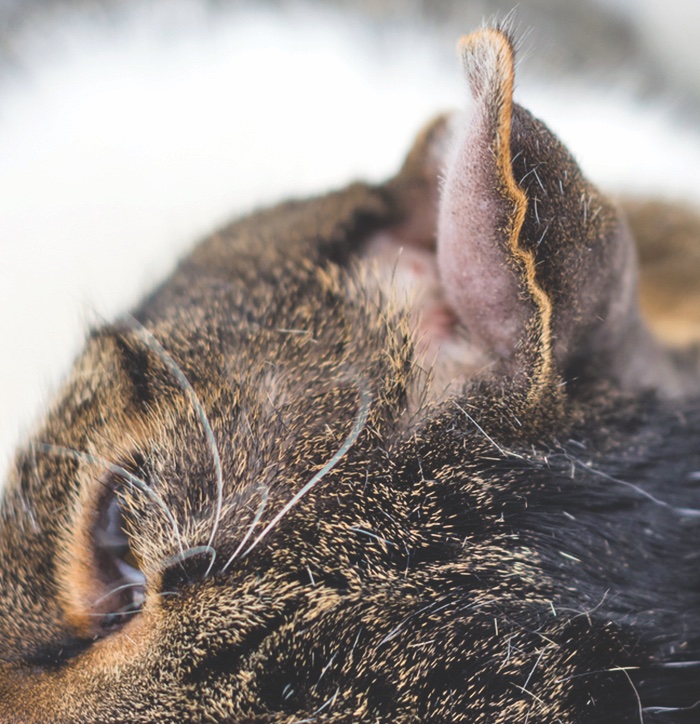
An aural hematoma occurs when blood pools under the skin on the flap of the ear. This can occur when one or more blood vessels on the ear rupture and bleed into the surrounding tissues.
A cat’s pinna (external ear) is composed primarily of layers cartilage and skin, and when blood accumulates between these layers, a hematoma can form on the ear.
As it forms, an aural hematoma causes a taut, fluid-filled balloon-like structure. It can feel warm to the touch, look discolored, and may be painful. The weight of the trapped blood generally causes the ear to droop, and you may notice your cat tilting her head due to the added pressure and weight.
What Causes an Aural Hematoma?
Any trauma that leads to the rupture of blood vessels in the pinna can cause an aural hematoma. A cat fight, head trauma, or self-induced trauma caused by your cat scratching at her ears because of ear mites can lead to a hematoma.
While not a get-to-the-clinic-immediately emergency, an aural hematoma should be treated as soon as possible. Your veterinarian may try using a needle to drain as much blood as possible. The affected ear may be taped onto the cat’s head to apply pressure in an effort to prevent the recurrence of any bleeding. Unfortunately, this technique is often ineffective, and the hematoma may redevelop over variable periods of time.
Surgery Is Best
Surgery is generally the best option, both cosmetically and for the future health of that ear.Luckily, this is a quick surgery that can be done at most clinics.
“We anesthetize the cat, make an incision on the inside of the pinna, drain the accumulated blood from the pinna, and then suture the inside and outside of the ear flap together, so that the inner skin is pressed flat against the outer skin. By doing this, the space where blood could accumulate in the future is obliterated,” says James Flanders, DVM, emeritus associate professor in the section of small animal surgery at Cornell University’s College of Veterinary Medicine.
With this technique, the sutures go all the way through the ear flap. Alternative versions may use a drain sutured on the inside of the ear to allow drainage until the injury naturally clots and scars over. Buttons also may be incorporated into the sutures to add pressure to the incision sites.
Post surgery, some cats will require a bandage to prevent them from scratching or rubbing at the surgery site. Some cats will require a cone to protect the area. Sutures can usually be removed in seven to 10 days. The scarring from the surgical procedure should prevent any future hematomas from developing in that ear.

If you decide not to treat a hematoma, your cat will usually end up with a “cauliflower ear” like human wrestlers sometimes get. As the clot disintegrates, it retracts and pulls the ear tissue with it. The resulting distorted ear is not only unsightly, but it may make your cat prone to more ear problems since the ear canal is closed up to varying degrees.


